Image Gallery: Capnography
Rachel Reed, DVM, DACVAA, University of Georgia
Capnometry involves the use of a capnometer to measure the amount of carbon dioxide (CO2) in expired gas. CO2 is produced as a by-product of metabolism in living cells and carried via the blood to the lungs, where it diffuses into alveolar gas and is expired. The amount of CO2 in expired gas is a direct representation of the adequacy of alveolar ventilation and an indirect indication of cardiac output and metabolic rate.
A normal end tidal CO2 (ETCO2) of 35 to 45 mm Hg is expected in the absence of hyper- or hypoventilation. Hyperventilation results in an abnormally low ETCO2 (<30 mm Hg), whereas hypoventilation results in an abnormally high ETCO2 (>50 mm Hg). A decrease in cardiac output or metabolic rate while alveolar ventilation remains unchanged results in a decrease in ETCO2. An increase in cardiac output or metabolic rate while alveolar ventilation remains unchanged results in an increase in ETCO2.
Related ArticlesTop 5 Anesthetic ComplicationsAnesthesia Monitoring: Raising the Standards of CareAnesthesia Monitoring & Pulse Oximeters
Capnography machines provide clinicians with a waveform (capnogram) created by expired CO2 as it is being measured by the machine. Understanding how to interpret capnograms can be useful in managing patients with hyper- and/or hypoventilation, along with identifying impending cardiac arrest and airway leaks, as well as identifying and troubleshooting complications associated with anesthetic equipment. Capnography arguably can provide more information about the status of patients than do other traditional methods of anesthesia monitoring.
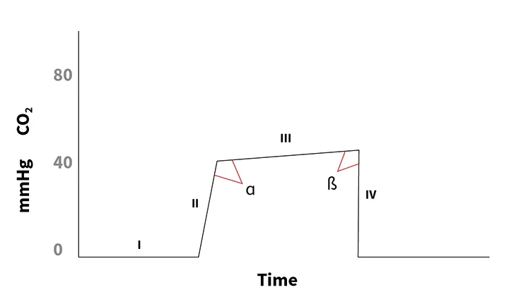
Figure 1 The four phases of capnography
Capnography has four phases as depicted on capnograms. Phase I is the inspiratory phase; during this time, the CO2 level should remain at zero. Phase II is the ascending phase represented by emptying of the trachea and bronchi and beginning of alveolar gas. Phase III is referred to as the alveolar plateau, which may appear flat but should have a gentle upward slope. ETCO2 is the peak at the end of the alveolar plateau. Phase IV is the descending phase represented by termination of expiration and beginning of inspiration as the CO2 level rapidly falls to zero. The angle between phases II and III, called the alpha angle, should be 100° to 110°. The angle between phases III and IV, called the beta angle, is normally ≈90°.