The Drooling Drunken Dog

Dale, a 4-year-old, 25-kg, castrated crossbreed dog, was presented for a 4-hour history of staggering, vomiting, and excessive drinking and urinating.
History
Dale had been normal before this event. He is allowed to go on off-leash walks in the woods, has access to his owners’ garage, has had no other previous medical conditions, and and was current on all vaccinations (including leptospirosis) vaccines.
Examination
Dale was quiet and slightly dull but responsive. Heart and lungs were normal on auscultation, with strong synchronous pulses. Vital signs were normal. The abdomen was tense on palpation, and he was drooling. A moderate-sized soft bladder was palpated, and the patient urinated a small amount during examination. Dale was ambulatory but ataxic and had normal general conscious proprioceptive reflexes and a normal cranial nerve examination. He was approximately 8% dehydrated. Body condition score was 5/9.
Multiple toxicities and disease processes can present with acute neurologic signs.
Diagnostics
Venous blood gas analysis (Table 1) showed metabolic acidosis with respiratory compensation and an increased anion gap (AG). A chemistry panel showed elevated blood urea nitrogen (BUN) and normal creatinine. The increased BUN was presumed to be because of dehydration. CBC showed hemoconcentration but was otherwise unremarkable. Free-catch urinalysis revealed isosthenuria and a significant number of calcium monohydrate crystals. Based on the suspicion of possible ethylene glycol (EG) toxicity from access to his owners’ garage (where antifreeze was present), a Kacey Ethylene Glycol Test Strip (Kacey Diagnostics) was performed and was positive for EG at a lethal dose of 50 mg/dL.
Table 1. Significant Laboratory Results
| CBC: Red blood cells (×106/µL)
| 9.2 | 5.83-8.87 | |
BLOOD CHEMISTRY: Blood urea nitrogen (mg/dL)
| 40 | 5-30 | |
BLOOD CHEMISTRY: Creatinine (mg/dL)
| 1.4 | 0.7-1.8 | |
VENOUS BLOOD GAS: pH
| 7.29 | 7.35-7.46 | |
VENOUS BLOOD GAS: Partial pressure CO2 (mm Hg)
| 22 | 40-46 | |
VENOUS BLOOD GAS: Bicarbonate (HCO3<sup–sup>; mmol/L)
| 12.8 | 22-24 | |
VENOUS BLOOD GAS: Chloride (mEq/L)
| 115.4 | 109-120 | |
VENOUS BLOOD GAS: Sodium (mEq/L)
| 160 | 140-150 | |
VENOUS BLOOD GAS: Potassium (mEq/L)
| 4.8 | 3.9-4.9 | |
VENOUS BLOOD GAS: Base excess (mmol/L)
| -10.2 | -4 - +4 | | URINALYSIS: Urine specific gravity | 1.010 | 1.018-1.050 | | URINALYSIS: Sediment | Calcium monohydrate crystals present | Negative | | ETHYLENE GLYCOL TEST: Kacey Ethylene Glycol Test Strips | Positive for 50 mg/dL | Negative |
ASK YOURSELF
What was Dale’s AG? What are the diagnostic differentials for an increased AG?
What is the toxic dose for EG in dogs vs cats?
In what stage did Dale present for EG toxicosis, and what are the stages of toxicity with EG?
What are current options for treatment for EG toxicosis and during what timeline do they need to be instituted in dogs as compared to cats?
Diagnosis
Ethylene glycol toxicity
Treatment
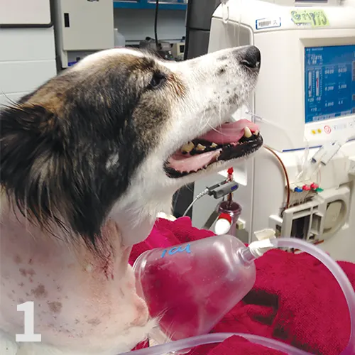
Patient receiving hemodialysis for EG toxicity.
Dale was hospitalized, and IV fluid therapy with a balanced electrolyte solution (PlasmaLyte A, 6 mL/kg/hr) was initiated. A bolus of 30% ethanol (1.31 mL/kg, slow IV) was administered, followed by 30% ethanol (0.42 mL/kg/hr CRI IV) as an antidote for EG toxicosis (Table 2). Dale was placed under general anesthesia, and a dialysis catheter was placed in the right jugular vein. He received an 8-hour hemodialysis treatment (Figure 1) with a urea reduction ratio goal of 95%. A high blood flow rate of 20 mL/kg/min was well-tolerated and a dialysate flow of 500 mL/min was used. The dialysate was formulated with 4 mmol/L potassium, 30 mmol/L bicarbonate, and 140 (mmol/L) sodium.
Ethanol was added to the dialysate concentration to achieve a dialysate ethanol concentration of 0.1%. Dale was also maintained on an IV ethanol infusion throughout the procedure.
Table 2. Suggested Protocols for Ethylene Glycol Intoxication
20% ethanol: 5.5 mL/kg IV every 4 hours for 5 treatments, then every 6 hours for 4 treatments or same dose administered as CRI until EG levels are negative, which is typically more than 48 hrs.
30% ethanol: 1.3 mL/kg slow IV bolus, then 0.42 mL/kg/hr CRI for 48 hours
|
20% ethanol: 5.0 mL/kg IV every 6 hours for 5 treatments, then every 8 hours for 4 treatments
30% ethanol: 1.3 mL/kg slow IV bolus, then 0.42 mL/kg/hr CRI for 48 hours
| |
4-METHYLPYRAZOLE (FOMEPIZOLE)
(50 mg/mL)*
|
Initial 20 mg/kg slow IV bolus over 15-20 minutes
At 12 and 24 hours after initial bolus, 15 mg/kg IV
At 36 hours after initial bolus, 5 mg/kg IV
|
Initial 125 mg/kg slow IV bolus
31.25 mg/kg IV every 12 hours after initial bolus for 3 treatments
|
*Antizol-Vet (fomepizole, Paladin Labs) has been withdrawn from the market, but human-use fomepizole and compounded versions are available.
Outcome
Dale did well during the hemodialysis treatment and was bright, alert, and eating normally the next day. On recheck bloodwork the following morning, his renal values, BUN, and venous blood gas parameters were normal. The patient was maintained on IV fluids for an additional 24 hours and was then discharged. On recheck 2 weeks later, Dale was doing well and renal values remained normal.
Related Article: Evaluating Ataxia in Suspected Ethylene Glycol Toxicity
DID YOU ANSWER?
The AG is used to help differentiate the causes of metabolic acidosis, which can be classified as an increased AG (with normochloremia) or a normal AG (with hyperchloremia). It is calculated as the difference between the measured cations and anions:Anion gap = ([Na<sup+sup>] + [K<sup+sup>]) – ([Cl<sup–sup>] + [HCO3<sup–sup>])Dale’s AG was 36.6 with a normal chloride concentration. Normal AG is typically 12 to 24 mEq/L in dogs and 13 to 27 mEq/L in cats. Differentials for an increased AG include diabetic ketoacidosis, uremia, EG intoxication, and lactic acidosis. Less common causes include ingestion of other toxicants (eg, salicylates [such as aspirin-containing compounds], methanol, ethanol).
EG is found in high concentrations in antifreeze, some windshield deicing solutions, and other industrial solvents. It can also be found in low concentrations in paint, printer cartridges, and household chemicals, although the levels in these products are generally so low that they are unlikely to result in toxicosis. EG toxicity can be fatal because of the small amount that needs to be ingested for a lethal dose. Outcome is associated with time elapsed between ingestion and treatment. The lethal dose for dogs is 4.4 to 6.6 mL/kg.1 Cats are significantly more sensitive to EG, with a lethal dose of 1.5 mL/kg.<sup2sup>
EG is rapidly absorbed from the GI tract and reaches peak levels 3 hours after ingestion.3 EG can cause significant CNS depression and GI irritation in Stage 1. CNS depression develops within 30 minutes of ingestion and often manifests as incoordination, ataxia, and “drunkenness.” During Stage 1, dogs may have profound polyuria and polydipsia secondary to the osmotic diuretic effects of EG.<sup4–6sup>In Stage 2, CNS signs may resolve by about 12 hours, and clinical signs may seem to resolve briefly; during this stage, however, dogs may become dehydrated and develop tachycardia and tachypnea secondary to the developing metabolic acidosis. In Stage 3 (36 to 72 hours after ingestion), signs of severe renal dysfunction may be observed in dogs. Patients may have large swollen kidneys, may be oliguric to anuric, and may quickly progress to profound lethargy, vomiting, seizures, coma, and death. Cats are more susceptible to EG toxicity, have a lower LD50, and progress through the stages much more rapidly than dogs.<sup3,7–9 sup> The enzyme alcohol dehydrogenase (ADH) converts EG into its toxic metabolites, including calcium oxalate crystals that develop throughout the body and cause secondary acute kidney injury (AKI). Treatment of EG exposure focuses on competitive inhibition of ADH to prevent the formation of these toxic metabolites, particularly glycoaldehyde and glycolic acid. If untreated, calcium oxalate monohydrate crystals can be seen within 4 to 6 hours of ingestion in dogs (3 hours in cats).7,10 AKI can develop within 24 to 72 hours and is often fatal once azotemia has developed.
Using a glycol test kit that detects EG concentrations >20 mg/dL can be useful to diagnose suspected EG toxicity in dogs. This test is reliable in cats; however, because of their significantly lower EG level for toxicity, the test may remain negative even with a toxic exposure. False-positive results can occur with propylene glycol, glycerol, ethanol, formaldehyde, and metaldehyde. Severe elevations of lactate dehydrogenase or lactic acid may also cause false-positive results.11 Definitive diagnosis of EG can be performed using gas chromatography, but this is expensive and not readily available.12 Ideally, a quantitative EG level, which can be performed at a veterinary diagnostic laboratory or human hospital, should be obtained for definitive diagnosis and monitoring of treatment.
Because EG is absorbed rapidly, induction of emesis is not indicated except within 2 hours of ingestion. As EG does not reliably bind to activated charcoal, further decontamination with charcoal is not warranted. Therapy focuses on prevention of metabolism and increasing speed of excretion of EG. Treatment with the antidote, 4-methylpyrazole (4-MP; fomepizole), or ethanol should be implemented as soon as possible (and ideally within 8 to 12 hours of exposure for best outcome). Hemodialysis is an effective treatment modality for EG.<sup8,13 sup> If instituted within the first 6 to 8 hours of exposure, hemodialysis can remove the water-soluble EG and its toxic metabolites from the blood and may be curative in a single treatment. If AKI occurs, hemodialysis may also be used to support the patient while waiting to see if renal function returns, which may take months.
Aggressive stabilization and supportive care are also extremely important in treating these patients. Because of the GI, central nervous system, and osmotic diuretic effects of EG, dehydration and potential hypovolemia secondary to concurrent vomiting and polyuria make IV fluid therapy an essential component of treatment. Close monitoring of the patient’s acid–base and volume status, cardiovascular stability, and urine output is imperative. The prognosis for patients with EG toxicity depends on the amount ingested and the time elapsed between exposure and institution of treatment. If dogs are treated within 12 hours and cats within 3 hours, outcomes are typically good.<sup5,7–9,14sup> However, even if antidote therapy is implemented, the prognosis is grave if the patient has become anuric.
Related Article: Ethylene Glycol Toxicosis in Dogs & Cats
ADH = alcohol dehydrogenase, AG = anion gap, AKI = acute kidney injury, BUN = blood urea nitrogen, EG = ethylene glycol, 4-MP = 4-methyl–pyrazole
ERIN MCGOWAN, VMD, is second-year emergency and critical care resident at University of Pennsylvania. A graduate of University of Pennsylvania, she completed her small animal rotating internship at Red Bank Veterinary Hospital in Tinton Falls, New Jersey.
LORI S. WADDELL, DVM, DACVECC, is adjunct assistant professor in the intensive care unit at University of Pennsylvania Matthew J. Ryan Veterinary Hospital. Her research interests include colloid osmotic pressure and coagulation in critically ill patients. She previously worked as emergency clinician in private practice and completed a residency in emergency medicine and critical care at University of Pennsylvania. Dr. Waddell, a graduate of Cornell University, completed an internship at Angell Memorial Animal Hospital in Boston.