In the Literature
Jondeau C, Gounon M, Bourguet A, Chahory S. Epidemiology and clinical significance of canine distichiasis: a retrospective study of 291 cases. Vet Ophthalmol. 2023:26(4):339-346. doi:10.1111/vop.13091
The Research …
Authors of this study aimed to describe the epidemiologic factors and clinical significance of canine distichiasis over a 10-year period. Dogs with distichiasis (n = 291) received a complete eye examination, including vision testing, pupillary light reflex evaluation, slit-lamp biomicroscopy, indirect ophthalmoscopy, tonometry, Schirmer tear test, and fluorescein staining. Dogs with additional eyelid abnormalities were excluded.
Distichia prevalence was 5.5%; median patient age at diagnosis was 3.9 years. Significant prevalence was noted in purebred (eg, American cocker spaniels, Cavalier King Charles spaniels, English cocker spaniels), brachycephalic (eg, English bulldogs, boxers), and short-haired dogs. Of note, distichia was an incidental finding in 86.5% of cases and was not associated with ocular disease or discomfort. In dogs with clinical signs, 39% exhibited corneal ulceration (superficial, 28.8%; deep stromal, 10.2%). Distichia was associated with ectopic cilia in 4.1% of dogs.
… The Takeaways
Key pearls to put into practice:
A Finhoff transilluminator or other bright, focal light source in a dimly lit room may help locate abnormal cilia in the absence of high magnification. The eye should be illuminated from the lateral canthus to the medial canthus across the eyelid margin, following the curve of the palpebral fissure. Distichiae that are yellow, cream, or white can be difficult to observe unless light is transmitted across the eyelid at the correct angle. In this author’s experience, shih tzus often exhibit irritation with distichia and concurrent corneal disease with corneal scarring, pigment, vascularization, and intermittent blepharospasm,1 while cocker spaniels may have large numbers of distichiae without obvious signs of discomfort or corneal pathology.
Small cilia forceps with a head loupe can be used to remove distichiae; however, this technique provides only temporary relief, as distichiae regrow within weeks. Distichiae that align with an associated corneal ulcer should be removed to improve the healing process. A soft contact lens may be applied to reduce corneal irritation during healing and improve comfort. An Elizabethan collar can help prevent self-trauma.
Many young dogs with nonhealing ulcers thought to be associated with distichiae or spontaneous, chronic corneal epithelial defects (ie, indolent corneal ulcer, boxer ulcer) have concurrent ectopic cilia that cause corneal frictional irritation (Figure).2 Ectopic cilia are challenging to identify without magnification (eg, head loupe, slit-lamp biomicroscope) but are usually located in the superior eyelid margin, are 2 to 4 mm from the meibomian gland orifices in the palpebral conjunctiva, and appear as small, red, raised swellings surrounded by a small conjunctival pigmented ring. Ectopic cilia can be surgically removed via excision with a punch biopsy; manual removal is typically not successful or possible.
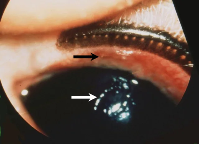
Ectopic cilia (black arrow) causing a nonhealing superficial corneal ulcer (white arrow) in a dog
Surgical intervention is needed for irritation from distichiae not caused by other eyelid abnormalities. In this author’s opinion, electroepilation and nitrous oxide cryosurgery are likely to improve patient comfort.3 Both procedures temporarily depigment the eyelid margins, cause transient eyelid swelling, and may alter tear dynamics.4 Topical tear film replacement may improve comfort and reduce corneal frictional irritation in brachycephalic dogs.
You are reading 2-Minute Takeaways, a research summary resource proudly presented by Clinician’s Brief. Clinician’s Brief does not conduct primary research.