Differentiating Exophthalmos, Buphthalmos, & Proptosis
Ron Ofri, DVM, PhD, DECVO, Hebrew University of Jerusalem

You have asked...
My patient’s eye appears abnormal. How do I know whether the eye is proptosed, exophthalmic, or buphthalmic?
The expert says...
Clinicians presented with patients whose eyes may appear asymmetrically sized must determine whether the irregular appearance is caused by proptosis, exophthalmos, or buphthalmos. The following guidelines can help differentiate these similar-appearing yet distinct conditions.
Related Article: Proptosis Reduction
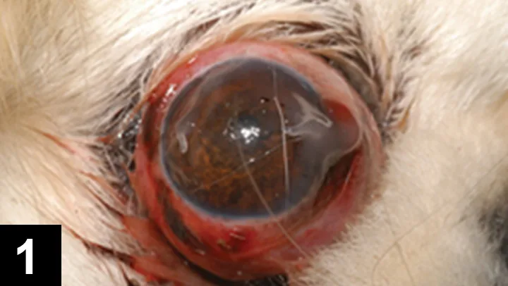
Figure 1. Traumatic proptosis of the eye in a Pekingese dog, a breed that is predisposed due to lagophthalmos and orbital confirmation. Note that the lower eyelid is now behind the equator of the globe. Conjunctival trauma and hemorrhage can also be seen. Lubricant has been applied to the cornea to prevent dessication. Courtesy Davis Veterinary Ophthalmology Service

Ocular Proptosis
With ocular proptosis (Figure 1), history often includes head trauma or a missing pet that returned with the condition. Careful examination reveals that the globe’s equator is visible and positioned anterior to the lids, preventing blinking. Stumps of torn extraocular muscles, strabismus, and intraocular injury may also be evident.
Exophthalmos & Buphthalmos
Exophthalmos involves a normal-sized globe that is pushed forward by a space-occupying lesion in the orbit, most commonly a retrobulbar abscess, cellulitis, or neoplasm. Buphthalmos, on the other hand, involves a normally positioned globe that is enlarged because of glaucoma. Despite differences in globe size and causes, however, clinicians may find it difficult to differentiate the two syndromes, as both involve red, asymmetric irregularity of the globe.
Exophthalmos and buphthalmos can often be differentiated during examination without additional instrumentation.
Some diagnostic procedures (eg, ultrasonography, tonometry) may provide a definitive diagnosis; although, exophthalmos and buphthalmos can often be differentiated during examination without additional instrumentation.
Clinicians should consider the following questions when differentiating between exophthalmos and buphthalmos (ie, glaucoma).
Related Article: Exophthalmos in a Dog
Is the condition unilateral or bilateral?
Glaucoma may be unilateral or bilateral, but exophthalmos is typically unilateral. Therefore, bilateral presentation usually indicates glaucoma.
How much conjunctiva is visible?
In exophthalmos, the eye is pushed forward; therefore, excess conjunctiva is visible, especially when looking at the eye from the side. With buphthalmos, the eye is stretched but remains in normal position inside the orbit; therefore, excess conjunctiva is usually not visible.
What is the position of the nictitans?
The nictitans (ie, third eyelid) is positioned normally in most glaucoma cases, although severe pain may sometimes cause enophthalmos and passive elevation of the nictitans. The nictitans is usually elevated in exophthalmos, as the space-occupying retrobulbar mass pushes against the nictitans, causing the elevation.
What is the diameter of the cornea?
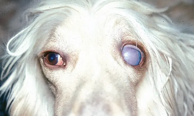
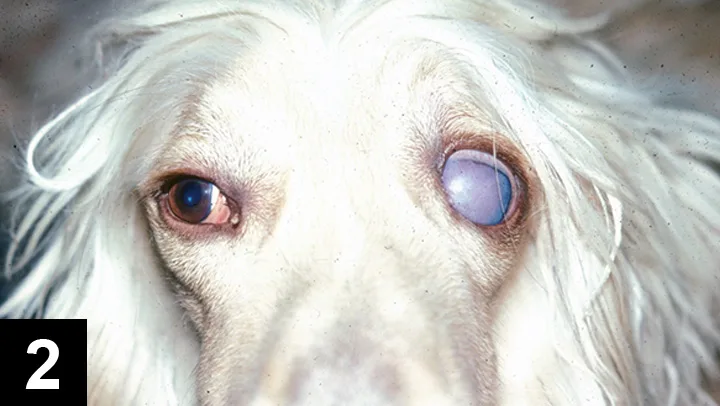
Figure 2. Unilateral glaucoma in an Afghan hound (4 years of age). Note the obvious difference in corneal diameter and corneal edema. Although vessels in the affected eye are more congested, the conjunctiva looks normal and the nictitans is not elevated, thus differentiating it from exophthalmus. Courtesy of Dr. Seth Koch

The corneal diameter is normal in exophthalmos and increased in buphthalmos because of globe stretching. This measurement can be sensitive; in many cases, even slight increases in corneal diameter can be detected, especially when comparing corneas with unilateral cases (Figure 2).
What are the results of a retropulsion test?
In a retropulsion test, two fingers are used to gently push on the globe through the upper eyelid. With buphthalmos, the eye may feel hard but will sink readily into the orbit. With exophthalmos, resistance to the retropulsion results from presence of a retrobulbar space-occupying mass. This test is not a measurement of intraocular pressure but of resistance.
What does the conjunctiva look like?
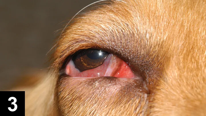
Figure 3. Exophthalmos caused by a retrobulbar mass in a golden retriever (2 years of age). Note that the lids are pushed forward, the nictitans is elevated, and the conjunctiva is congested. However, no corneal edema is evident, nor were there signs of intraocular disease, thus differentiating it from glaucoma. Courtesy of Dr. David Maggs

Glaucoma may present as a red ciliary flush (ie, redness more pronounced near the cornea, or a red ring around cornea), congested episcleral vessels, slightly congested conjunctival capillaries, and lacrimation, but the conjunctiva itself is normal in consistency. Depending on the cause, exophthalmos may present with chemosis (the retrobulbar mass impedes orbital circulation), and signs of conjunctivitis and purulent discharge (in cases of abscesses) may be present (Figure 3).
Is there evidence of pain when opening the mouth?
Glaucoma, retrobulbar abscesses, and retrobulbar tumors are all potentially painful; however, if exophthalmos is caused by a retrobulbar abscess, an increased pain response may be evident when the clinician attempts to open the patient’s mouth (opening the mouth causes the ramus of the mandible to press against the abscess). The patient may thus vocalize or struggle. With glaucoma, opening the mouth will not affect the pain response.
Are there any unique signs?
Corneal striae (ie, linear streaks seen in the cornea because of breaks in Descemet’s membrane caused by buphthalmos and corneal stretching) and corneal edema are associated with glaucoma, but not with exophthalmos. In contrast, mandibular lymph nodes may be enlarged in many cases of exophthalmos (caused by either a retrobulbar tumor or abscess) but will be normal in size for glaucoma. Vision and the presence of a pupillary light reflex are not reliable differentials, as optic nerve damage may occur with either condition.
Further Diagnostics
The previously noted clinical observations are often sufficient for a tentative diagnosis. However, two procedures that do require instrumentation can provide a conclusive answer.
Tonometry
Intraocular pressure will be normal in patients with exophthalmos, in contrast to the elevated pressure that occurs with glaucoma.
Imaging
Ultrasonography is useful for imaging retrobulbar masses in cases of exophthalmos. Ultrasonography can also be used to measure the axial length of the globe, helping to determine whether it is normal or enlarged. Advanced imaging (eg, CT, MRI) may yield additional diagnostic information regarding exophthalmos, as would ultrasound-guided fine-needle aspiration.
Conclusion
Asymmetric presentation of one or both eyes requires careful examination to determine whether the presentation is caused by traumatic proptosis, glaucoma, or retrobulbar disease. Achieving a definitive diagnosis is critical, as there are differences in causes, treatment, and long-term prognosis.