Conjunctivitis in Cats
Ron Ofri, DVM, PhD, DECVO, Hebrew University of Jerusalem
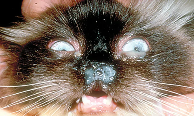
Rhinosinusitis, conjunctivitis, and mucopurulent ocular discharge in a patient with FHV-1 infection. Photo courtesy of Michael G. Davidson, DVM, DACVO
Profile
Definition
Conjunctivitis is an inflammation of the conjunctiva, a tissue that lines the eyelids and covers the sclerae.
The conjunctiva is an exposed mucous membrane that reacts to antigenic stimulation caused by contact with noxious stimuli.
The superficial stroma of the conjunctiva is rich in lymphatic tissue, both diffuse and aggregated.
When aggregated tissue is stimulated, it forms lymphoid follicles, which produce effector cells. Conjunctival plasma cells produce specific immunoglobulins as part of the secretory component of the immune response.1,2
Causes
Infectious primary pathogens
Cause of most cases of feline conjunctivitis1,3-6
Feline herpesvirus type 1 (FHV-1)
Most common cause of conjunctivitis in cats
Studies suggest that 95% of cats worldwide have been exposed to the virus, and at least 80% of cats are latent carriers of the virus.7-11
Chlamydophila felis
A common cause of conjunctivitis, especially in young kittens6,12-14
Calicivirus
Primarily a respiratory tract pathogen that may cause mild and transient conjunctivitis
Most cats will recover spontaneously.15
Mycoplasma spp
Can be present in healthy cats, however, and may thrive because of coinfection with FHV-1 or C felis, making its clinical significance questionable1,3-6,12-13
Because of their epidemiologic prevalence, only FHV-1 and C felis will be discussed in this article.
Pathophysiology
FHV-1 conjunctivitis
The primary disease commonly occurs in kittens and is caused by exposure to wild-type FHV-1, which is transmitted between cats by microdroplets (frequently from the queen) or fomites (possibly by the client).1,3
FHV-1 infection is characterized by conjunctivitis, respiratory tract signs (Figure 1), and, less frequently, corneal ulceration.4,5
Following primary, and usually self-limiting, disease, FHV-1 establishes lifelong latency in the trigeminal ganglia.
The virus is cleared in only a limited number of cats.7
Stress or treatment with steroids will induce subsequent reactivation and shedding of the virus and may result in recrudescent disease.8-9
Recrudescent disease is usually milder than the primary infection and can affect the cornea, conjunctiva, and/or respiratory system.9,11,16
The recurrent disease may be cytolytic (because of viral replication) and cause conjunctival (Figure 2) or corneal (Figure 3) ulceration; it also may be immune- mediated and cause stromal keratitis (Figure 4) or chronic lymphoplasmacytic conjunctivitis.1,4,11
C felis conjunctivitis
Cats are infected by airborne transmission or contact with infected cats or fomites.1,3
The infection initially may be unilateral.
It usually spreads to the other eye within a week, but some cases may remain unilateral.4
If untreated, chronic disease, characterized by membranous or follicular conjunctivitis, may occur, or the cat may become a subclinical carrier and contribute to the spread of the disease.5
Natural immunity may develop, and the disease is rarely seen in cats older than 5 years of age.17
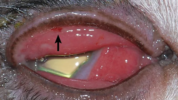
Figure 2
Conjunctivitis and conjunctival ulceration (black arrow) in a patient with FHV-1 infection. Although C felis may cause conjunctivitis, presence of an ulcer indicates a viral disease. Photo courtesy of Michael G. Davidson, DVM, DACVO
History & Physical Examination
In cases of FHV-1 conjunctivitis, client questioning may reveal stressful events (eg, rehousing, traveling, introduction of a new pet or baby to the household) preceding the appearance of clinical signs.
Pregnancy, parturition, lactation, concurrent illness, or treatment with glucocorticoids can also induce viral shedding and recrudescent disease.1,3-5,7,17
In kittens with FHV-1 conjunctivitis, physical examination may show severe signs of upper respiratory disease, including fever, sneezing, rhinitis, and purulent nasal discharge.
These signs are milder in cases of C felis conjunctivitis and in adult cats with FHV-1 conjunctivitis and may include nasal discharge and sneezing.1,3-5,11,16
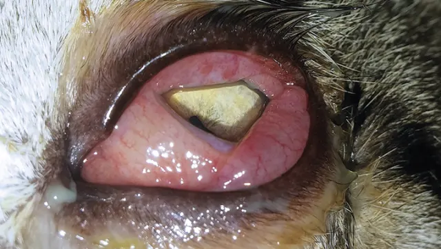
Chemosis and conjunctivitis without corneal involvement in a patient with C felis infection
Clinical Signs
Clinical signs are usually more severe in FHV-1 primary disease in kittens and milder in C felis and adult FHV-1 disease.
Clinical signs may help distinguish between the pathogens.1,3-5,7,11,16,17
Hyperemia of conjunctival vessels (ie, red eye) is usually more marked in FHV-1 conjunctivitis than in C felis conjunctivitis.
Conjunctival edema (chemosis), swelling, and thickening is usually more marked in C felis conjunctivitis than in FHV-1 conjunctivitis (Figure 5).
Ocular discharge may be mucoid in C felis conjunctivitis and adult FHV-1 conjunctivitis and purulent in kittens with FHV-1 conjunctivitis (Figure 1).
Conjunctival ulceration is more characteristic of FHV-1 disease, especially in kittens (Figure 2).
Concurrent corneal involvement and ulceration may be seen in FHV-1 conjunctivitis but not in C felis conjunctivitis (Figures 3 and 4).
Minimal ocular pain, but possible discomfort, expressed as blepharospasm, may be seen in conjunctivitis but is marked in cases of corneal ulceration.
Diagnosis
Clinical Diagnosis
As compared with C felis infection, FHV-1 infection causes greater conjunctival hyperemia and ocular discharge, and respiratory signs are usually more severe, especially during the primary disease (Figure 1).
Recurrent disease, keratitis, and corneal and conjunctival ulceration are definitive hallmarks of FHV-1 infection (Figures 2-4).
Presence of symblepharon, or adhesions between the cornea and conjunctiva, indicates previous FHV-1 infection.1,3-5,7,17
Chemosis is more severe in C felis infection than in FHV-1 infection and is the most notable clinical sign (Figure 5).
Although infection with C felis is generally mild, chlamydial conjunctivitis can be persistent, especially if not treated.
In chronic stages, membranous or follicular conjunctivitis may develop.1,3-5,7-8
Diagnosis may also be based on response to treatment.
FHV-1 involvement should always be suspected in patients with conjunctivitis.
C felis infection may be considered in patients with acute chemosis or chronic disease.1,4,11
Differential Diagnoses
Eyelid and eyelash disorders
Rare causes of conjunctivitis in cats
Dry eye (ie, keratoconjunctivitis sicca) is commonly a sequela of FHV-1 infection rather than a primary cause of feline conjunctivitis.1,4,11
Calicivirus, Mycoplasma spp, and Bordetella bronchiseptica1,4,12-13,15
Lipogranulomatous, eosinophilic, and parasitic conjunctivitis1,3-5,17
Red eye
Clinical sign of anterior uveitis and glaucoma
Appropriate testing for these conditions should be performed.
Laboratory Findings
Diagnostic tests for FHV-1 include immunofluorescent antibody testing, viral isolation of FHV-1 in feline cell cultures, and polymerase chain reaction (PCR).1,3,4
False-negative and false-positive test results are common because of subclinical shedding of FHV-1 by healthy animals, reduced shedding in recrudescent stages of the disease, and high prevalence of antibodies from vaccination and exposure.12-14
Diagnostic tests for C felis include PCR and cytologic evaluation. Presence of intracytoplasmic elementary bodies in epithelial cells collected from conjunctival scrapes (Figure 6) is indicative of C felis infection.12-14,18
These bodies are transient and can be difficult to identify.
As the utility of diagnostic testing is limited, treatment based on clinical signs without confirmation of the underlying disease is an acceptable approach to conjunctivitis in cats.1,3-5,7,11
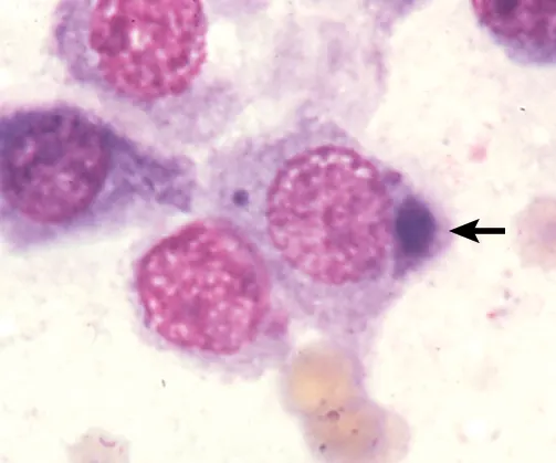
Presence of an intracytoplasmic elementary body in an epithelial cell collected from a conjunctival scrape (arrow) is indicative of C felis infection. Photo courtesy of Michael G. Davidson, DVM, DACVO
Treatment
Inpatient or Outpatient
Reducing stress is important in treating FHV-1 infection and may help determine the choice of therapy.
Some topical antiviral drugs require frequent administration, which can increase patient stress. In mild cases, no treatment may be preferable.1,3-5,7,11,17
When possible, patients with conjunctivitis should receive treatment at home, where clients can provide a nurturing and less stressful environment.
Medical
Medical management includes use of topical and oral antivirals and antibiotics.
C felis conjunctivitis
Treated with topical ointment containing 1% tetracycline q6-8h for 1 to 2 weeks
Recent studies suggest that topical treatment be supplemented, or even replaced, with oral doxycycline 10 mg/kg q24h for 3 weeks.
Systemic treatment may be indicated in patients with concurrent respiratory disease.
Systemic treatment is also useful in preventing secondary ocular bacterial infection in FHV-1 conjunctivitis.19,20
Rapid resolution of signs during treatment may suggest that C felis is the causal agent.
Topical antiviral drugs
Usually administered 5 to 6 times a day, with treatment continuing for 10 to 14 days after resolution of signs21,22,25
Trifluridine 1%, idoxuridine 0.1%, and vidarabine 3%
Variably effective19,21-25
Trifluridine has the highest efficacy and provides transcorneal penetration but may be more irritating to cats.19-23
Idoxuridine and vidarabine are less irritating but may be difficult to obtain because they are not widely available commercially.
They can be ordered from compounding pharmacies.
Cidofovir 0.5%
Unavailable commercially as an ophthalmic preparation
Has strong in vitro and in vivo efficacy against FHV-1 infection, with treatment reducing severity of clinical signs and viral shedding26-28
Has beneficial effects when administered q12h, which is a significant advantage as compared with other topical antiviral medications.26-28
Less toxic than other antivirals because of its relatively high specificity for viral—rather than host—replication proteins26-28
Systemic antiviral treatment
Famciclovir
A prodrug of penciclovir
Safe and effective for treating FHV-1 conjunctivitis
Recommended dose is 90 mg/kg q12h29-33
All current systemic and topical antiviral drugs are virustatic and achieve their effect by interfering with active viral DNA replication.
They are ineffective at eradicating latent infection.
Significant toxicity can occur with anti-viral administration because of the intracellular location of the virus and the inability of available medications to selectively target viral—rather than host cell—replication.19,21-25
Many commercially available antiviral drugs, notably acyclovir, are effective against human herpes simplex virus but ineffective against FHV-1.20-25
Others, such as valacyclovir, may be toxic to cats and should not be used.19,23
Most patients greatly benefit from frequent application of high-quality artificial tear (eg, hyaluronate) preparations, as FHV-1 infection reduces conjunctival goblet cells and causes qualitative tear film disorders.1,4,34,35
Glucocorticoid use in cats should be considered carefully, as these drugs may also induce viral shedding.
When glucocorticoid treatment is unavoidable (eg, in patients with eosinophilic keratitis or anterior uveitis), concurrent antiviral treatment should be provided and the patient monitored closely for recrudescent disease.
Because FHV-1 infection may be reacti- vated during immunosuppression, the prognosis is poor in immunosuppressed patients (eg, those with feline leukemia virus or feline immunodeficiency virus).1,3-5,7,11,17
Client Education
Clients should be advised that:
Antiviral drugs are ineffective against latent FHV-1 infection.
Few cats will clear the virus, and it will remain latent in most patients.
Even after clinical signs have resolved, recurrence of disease is possible, especially when cats experience stress.8,11,19
Follow-up & Complications
Treatment should be continued for 2 weeks after resolution of clinical signs and the patient re-examined.8,19,23
FHV-1 has been implicated in the pathogenesis of corneal sequestrum, eosinophilic keratitis, and dry eye.4,7,17
These should be regarded as possible sequelae of infection.
In kittens, FHV-1 keratoconjunctivitis may result in ulceration of both the cornea and conjunctiva.
These ulcerated tissues may adhere to each other and form symblepharon (Figure 7), which is challenging to treat and requires surgical intervention.4,7,17
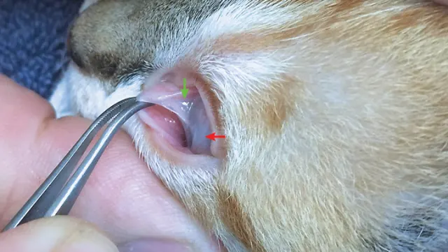
Symblepharon, or adhesions between the cornea and conjunctiva, is a common complication of FHV-1 keratoconjunctivitis. In this case, most of the cornea is obscured because of adhesions of the bulbar conjunctiva (red arrow). Adhesions between the bulbar conjunctiva of the third eyelid and cornea (green arrow) are also present.
Prognosis
Most patients with FHV-1 infection will remain carriers of the virus.
Recurrence is possible, particularly for patients in a stressful environment.
Cats also may be subclinical carriers of C felis.
The carrier state is not characterized by recurrent disease, but it contributes to the spread of C felis infection to other cats.1,4,13,15