Cecal Perforation During Ovariohysterectomy in a Rabbit
A young female rabbit was presented for elective ovariohysterectomy.
History
This rabbit had been relinquished to a rescue group 1 week before presentation and had displayed no signs of illness since her entry into foster care.
Examination
At presentation, the rabbit was bright and alert, the mucous membranes were pink with normal capillary refill time, and she appeared well hydrated. Findings on thoracic auscultation, abdominal palpation, and oral examination were within normal limits. Preoperative diagnostic testing was not performed due to financial limitations.
Anesthesia
Glycopyrrolate (0.005 mg/kg), midazolam (0.3 mg/kg), terbutaline (0.1 mg/kg), and hydromorphone (0.05 mg/kg) were administered subcutaneously approximately 30 minutes before anesthesia induction. An intravenous catheter was placed in the right cephalic vein, and anesthesia was induced intravenously with ketamine (2.8 mg/kg) and midazolam (0.15 mg/kg), after which the patient was intubated and maintained on isoflurane gas.
Monitoring of body temperature, oxygen saturation, and end-tidal carbon dioxide; electrocardiography; and assessment of indirect systemic arterial blood pressure with Doppler ultrasonography were performed throughout the anesthetic period. Adequate systolic blood pressure was maintained during the procedure by using a dopamine constant-rate infusion (3-7 mcg/kg per minute) as well as a slow hetastarch bolus (5 mL/kg). A bupivicaine (2 mg/kg) subcutaneous line block was performed along the ventral midline incision area before surgical incision. No anesthetic complications occurred during the procedure, and recovery was uneventful.
Procedure
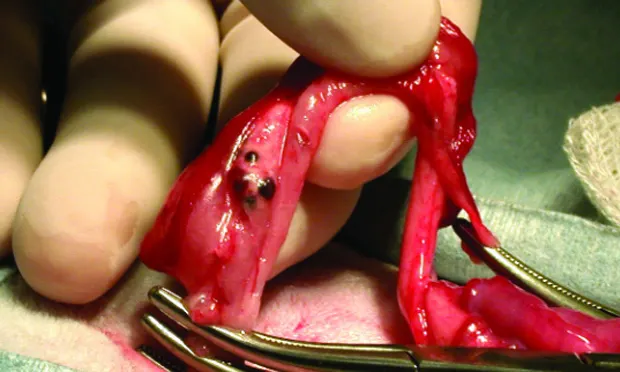
The routine ovariohysterectomy had been nearly completed when a full-thickness linear perforation about 8 mm long located on the ventral aspect of the cecum was noticed. The cecum was immediately isolated with laparotomy sponges, and the ovariohysterectomy was completed. No gross leakage of gastrointestinal contents was noted in the abdomen.
The cecal defect was repaired by using 5-0 absorbable monofilament glycomer 631 sutures (Biosyn; syneture.com) in a simple continuous pattern, and the abdomen was thoroughly lavaged with warm saline. A 2 ¥ 4-cm piece of oxidized regenerated cellulose (Surgicel mesh; surgicel.com) was placed over the repaired cecal defect before closure.
ASK YOURSELFWhich of the following are essential considerations in developing the postoperative management plan for this case?A. Postoperative pain managementB. Broad-spectrum antibiotic therapyC. Management of ileusD. Prevention of postoperative adhesion formationE. All of the above
CORRECT ANSWER E:All of the Above
Management of ileus (to which postoperative pain and stress contribute) and peritonitis is essential to successful immediate recovery in cases such as this, and prevention of adhesions will prevent future gastrointestinal motility problems and pain.
Management
Antibiotic therapy with cefoxitin (30 mg/kg IV Q 90 minutes, 2 doses) was initiated during surgery as soon as the cecal defect was identified. The rabbit recovered uneventfully from anesthesia. The animal's body temperature, heart rate, respiratory rate, pain score, body weight, packed cell volume and total solids, and arterial blood gas and electrolytes were closely monitored over the next 24 hours. All values remained within normal limits.
Image, left. Regular and frequent syringe feedings are a mainstay of ileus therapy; they provide oral hydration and nutrition, and help stimulate gastrointestinal motility.

The rabbit's pain and postoperative inflammation were controlled with a hydromorphone CRI (0.005-0.02 mg/kg per hour) and meloxicam (0.5 mg/kg SC Q 12 H). Antibiotic therapy initiated at anesthetic recovery included enro-floxacin (20 mg/kg IV Q 24 H) and metronida-zole (10 mg/kg IV Q 12 H), as well as continued dosing of cefoxitin (administered during surgery and 6 hours after recovery).
Postoperative gastrointestinal hypomotility was initially managed with a metoclopramide CRI (4 mg/kg Q 24 H) and fluid therapy (Normosol-R [hospira.com] plus 20 mEq/L of potassium chloride; 130 mL/kg Q 24 H). Syringe feeding of Oxbow Critical Care (oxbowanimalhealth.com) at 20 mL/kg Q 4 to 6 H was initiated the following morning.
Outcome
By 36 hours after surgery, the rabbit was passing normal fecal pellets and was removed from the critical care unit. She was switched to enrofloxacin (20 mg/kg PO Q 24 H for 10 days), metronidazole (10 mg/kg PO Q 12 H for 10 days), and meloxicam (0.5 mg/kg PO Q 12 H for 5 days). Supportive care for ileus was continued until the following day, when the rabbit displayed normal fecal production and hay consumption.
The rabbit's postoperative recovery continued smoothly, and she was released to the shelter 3 days after her initial presentation. One week after surgery the rabbit was reported to be doing very well, with no signs of infection or gastrointestinal dysfunction.
Discussion
Ovariohysterectomy should be recommended for any female rabbit not intended for breeding, primarily to prevent reproduction and highly prevalent uterine adenocarcinoma.1 The thin-walled cecum is the largest organ in the abdominal cavity of rabbits, containing approximately 40% of the gastrointestinal tract's ingesta for fermentation.2
During a ventral midline approach to the abdomen, it is necessary to use caution when entering the abdominal cavity because the cecum may be directly beneath the incision.2 In this particular case, it is unknown at what point the cecal wall was compromised (at the initial abdominal incision or during surgery) or how it was damaged (scalpel blade vs suture needle). Prompt and appropriate action was taken as soon as the defect was noticed, however, and the timeliness of this response contributed to the successful outcome. The initiation of broad-spectrum antibiotic therapy and intensive management of postoperative ileus were also integral to this rabbit's recovery.
Rabbits are used extensively as experimental models for the formation of intraabdominal adhesions,2 and adhesion formation is a significant concern whenever the gastrointestinal tract is manipulated. Many products have been investigated for the prevention of adhesion formation; in this case, surgical mesh (oxidized regenerated cellulose) was placed over the cecal repair as an absorbable adhesion barrier. In humans, the oxidized regenerated cellulose undergoes hydrolysis into a gelatinous protective barrier with antibacterial properties within 8 hours after application; it is completely absorbed within 28 days.3
Prognosis
The long-term effects of gastrointestinal perforation, repair, and possible intraabdominal adhesion formation have not been followed in this patient, and it remains uncertain how these factors will affect this rabbit's long-term prognosis. However, an 8-week postoperative update reports that she appears to have fully recovered, with no evidence of gastrointestinal complications.
Take-Home Message
Ovariohysterectomy should be strongly recommended in female rabbits not intended for breeding due to the high prevalence of uterine adenocarcinoma in intact animals.
Care must be taken when entering and working within the rabbit abdomen to prevent iatrogenic damage to the large, thin-walled cecum.
Intraabdominal adhesion formation is a significant concern whenever the rabbit gastrointestinal tract is manipulated.
Immediate recognition, broad-spectrum antibiotic coverage, and ileus management are key considerations in the postoperative care of cecal perforation.