Blood Smear Platelet Evaluation & Interpretation
Lisa M. Pohlman, DVM, MS, DACVP, Kansas State University

In most laboratories and veterinary practices, platelet concentration is determined via automated analysis. However, there are several variables that can affect accuracy of results, the most common of which, in the author’s experience, is platelet clumping. Platelet clumps must be identified on a blood film to avoid potential misdiagnosis of thrombocytopenia.
Analyzers that use impedance methodology can also produce erroneous results when platelet size overlaps with RBC size, causing large platelets to be counted as RBCs; this can result in a falsely decreased platelet concentration and misdiagnosis. This can occur in any animal that produces large platelets but is particularly common in cats, as their platelet size is similar to their RBC size.1,2
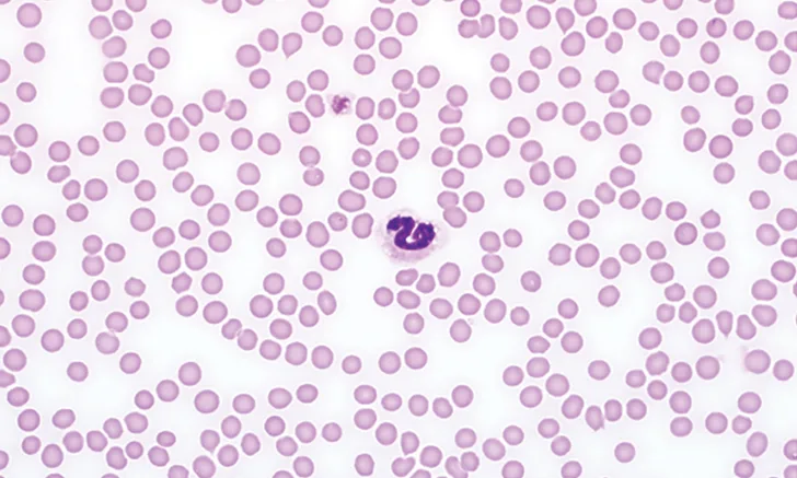
Estimation of the platelet concentration from a blood film should be performed in the monolayer using 100× objective (ie, 1000× magnification). A well made blood film with even distribution of platelets throughout the monolayer is essential. If platelet clumps are present, the platelet estimation will be falsely decreased to an unknown degree, depending on the amount of clumping. However, estimation using the method provided should help to provide the minimum concentration of platelets present.
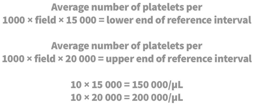
First, the entire film should be examined for clumps. Then, at least 10 fields within the monolayer should be reviewed to determine the average number of platelets per 1000× magnification field. The number observed should be multiplied by 15 000 to get the lower end of the reference interval and then by 20 000 to determine the upper end of the reference interval.2
